deep learning

Deep learning (nnUNet) model for the EPTN contouring guidelines for OARs in neuro-oncology
Ana M. Barragán-Montero, Margerie Huet-Dastarac, Dario di Perri, David Hofstede, Nikolina E Birimac, Benjamin Roberfroid, Emilien Quéré, John Lee, Wouter Van Elmpt, Danielle BP Eekers, Catharina M.L.Zegers


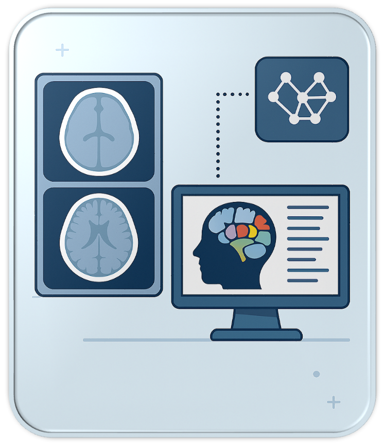
A representation of a model trained on neurological images to form a contouring atlas

The European Particle Therapy Network (EPTN) has undertaken a major effort to harmonize OAR contouring practices in neuro-oncology. In 2018, EPTN published the first consensus-based guidelines for the delineation of 15 brain OARs (10.17195/candat.2017.08.1), expanded in 2021 to include 10 additional structures (10.17195/candat.2021.02.1). To aid adoption, EPTN also produced explanatory videos (10.17195/candat.2022.02.1), but the broad implementation of these guidelines remains limited due to the complexity and time-consuming contouring. In this context, deep learning (DL) offers a promising solution. Expert-trained DL models can encode complex anatomical knowledge and replicate expert-level contouring.

To this end, an nnUNet architecture has been trained and made public here. The model was trained using a 5-fold cross validation on 74 patients treated at Maastro Clinic, contoured according to the EPTN guidelines. The model receives as input a planning CT scan with intravenous iodine contrast, along with 3T MRI scan gadolinium-enhanced T1-weighted sequence. CT and MR image should be rigidly registered. The output of the model are the 25 OARs defined in the EPTN consensus guidelines.
Deep Learning for Automated Ventricle and Periventricular Space Segmentation on CT and T1CE MRI in Neuro-Oncology Patients

Mart Wubbels, Marvin Ribeiro, Jelmer M. Wolterink, Wouter van Elmpt, Inge Compter, David Hofstede, Nikolina E. Birimac, Femke Vaassen, Kati Palmgren, Hendrik H. G. Hansen, Hiska L. van der Weide, Charlotte L. Brouwer, Miranda C. A. Kramer, Daniëlle B. P. Eekers, Catharina M. L. Zegers

Wubbels at al. developed and validate a deep learning model to automatically segment the ventricles and periventricular space on CT and MRI scans to improve treatment planning for patients receiving intracranial radiotherapy. The resulting model (based on nnU-Net) was tested alongside an existing model (SynthSeg) to see which performed better at segmenting the brain ventricles.

The results showed that the new nnU-Net-based model, performed more accurately and was preferred by radiotherapy technicians. These findings could improve the process of contouring organs at risk in brain cancer patients undergoing radiation therapy. To aid the use of the developed model, we provide additional documentation/code of the model which can be found in this repository: nnU-Net Ventricle Segmentation (GitLab).