Under Construction | This is a new version of CancerData. Please be patient while we update our content.
Welcome to CancerData
CancerData is a website that promotes free and open-source resource sharing.
We strongly believe that this drives research forward and benefits everyone. CancerData: together we care.
Research Focus
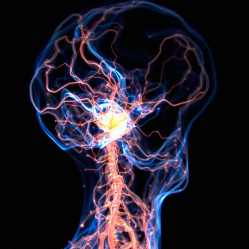
Neuro-Oncology
Building on expert knowledge
Years of international collaboration comes together in atlases and guidelines for contouring critical structures and its tolerance doses in the brain.Atlases: contouring the brain
Contouring brain structures in a uniform manner is essential for valuable, standardised data collection.
Constraints guidelines
Standardising constraints on critical organs helps in the generation of treatment plans with the best quality.