· David Hofstede , Dario Di Perri , Erik Roelofs , Alida Postma , Esther Troost , Daniëlle Eekers · Tutorial · 10.17195/candat.2022.02.1
Periventricular Space
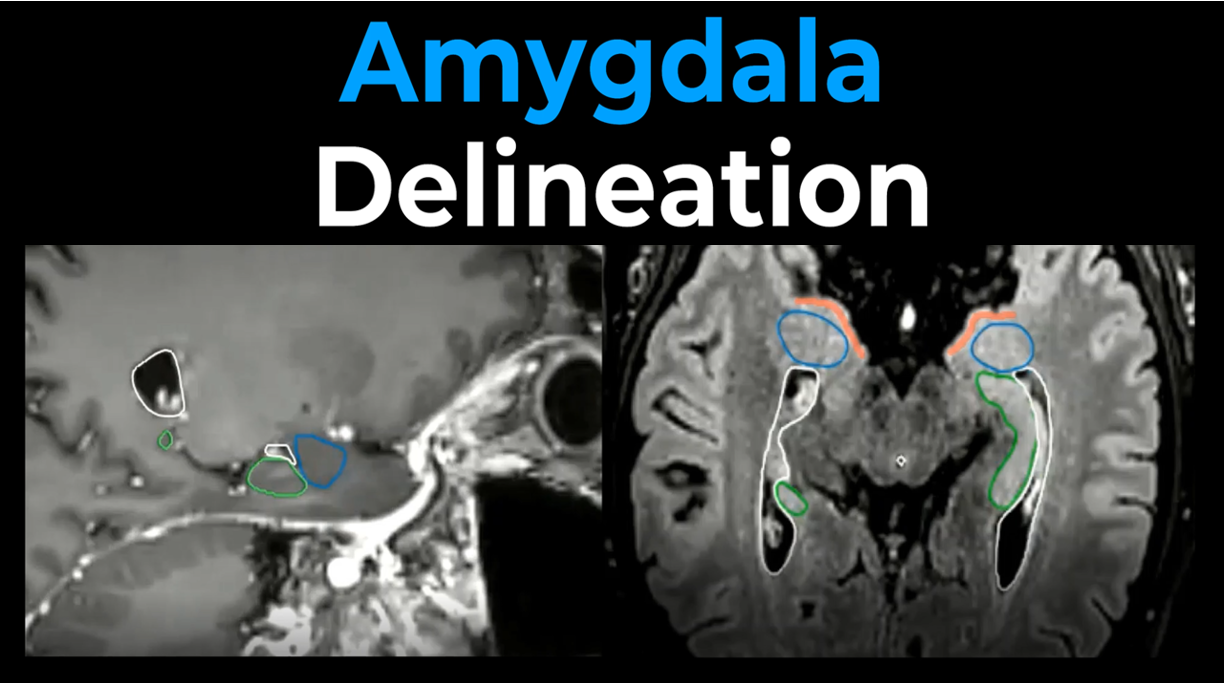
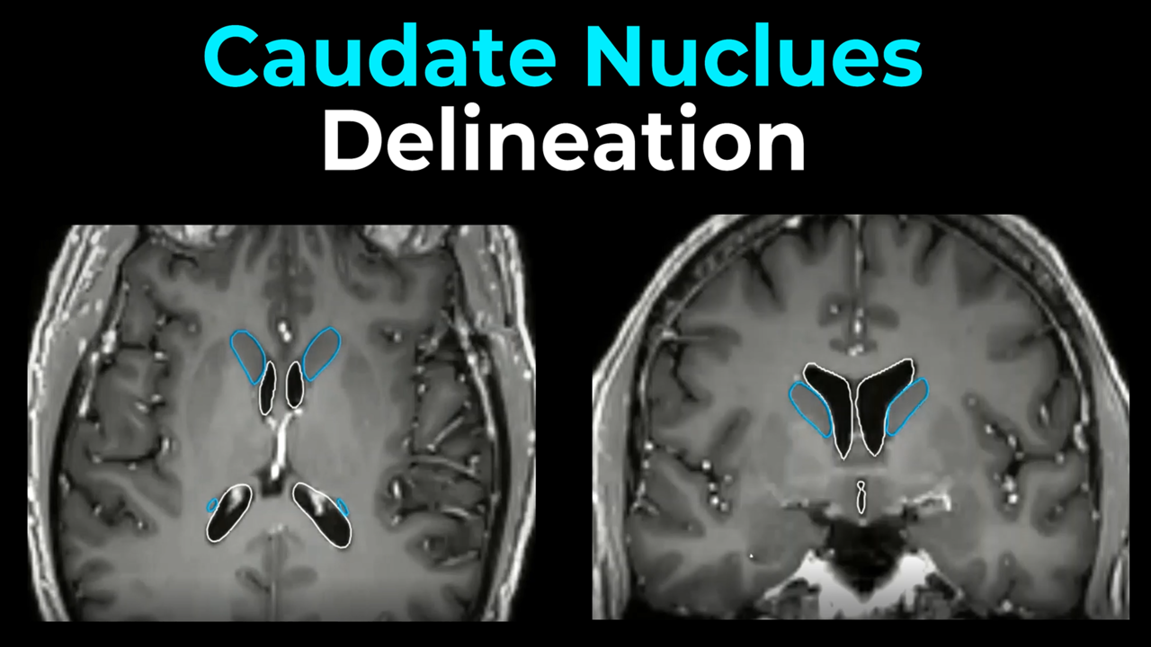
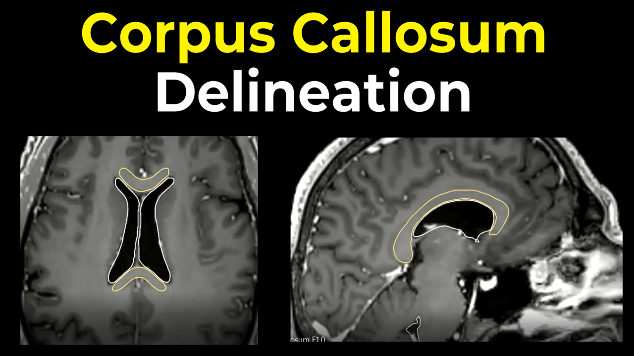
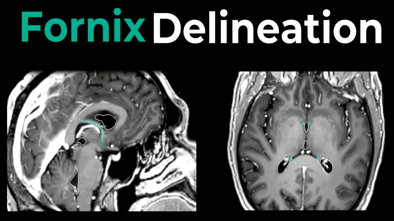
Contouring instructions for the periventricular space.
Periventricular Space
The periventricular space (PVS) comprises the brain tissue immediately surrounding the ventricular system. Growing evidence suggests that this region may be particularly susceptible to the development of late radiation-induced contrast-enhancing lesions following cranial radiotherapy. Several factors may contribute to this increased vulnerability, including the relatively limited vascular supply of periventricular tissues and, in particle therapy, potential variations in relative biological effectiveness (RBE) at the end of the beam range. The innermost portion of the PVS contains the subventricular zone (SVZ), one of the brain’s principal neural stem cell niches. The SVZ has attracted considerable interest because of its role in neurogenesis and its potential involvement in glioma development, progression, and treatment response. As a result, accurate delineation of the periventricular region has become increasingly relevant for both toxicity assessment and research into the biological effects of radiation on normal brain tissue and neural stem cell compartments.
Co-registration with a T1-weighted MRI is recommended to facilitate accurate delineation. In cases where ventricular margins are difficult to identify, co-registration with T2-weighted MRI may provide additional anatomical detail. The PVS can be defined as a 5 mm expansion around the ventricular system, including all ventricular compartments and their anatomical connections.
The ventricular system consists of:
- the lateral ventricles
- the third ventricle
- the fourth ventricle
- the ventricular connections
The size and configuration of the ventricular system can vary considerably between patients. In some cases, particularly in the presence of small or partially collapsed ventricles, certain ventricular components can be difficult to identify. Careful review of all available imaging sequences is therefore recommended to ensure accurate and consistent delineation.
The accompanying video demonstrates a step-by-step approach for generating and reviewing the periventricular space contour using the ventricular system as the anatomical reference. This educational resource is intended to support consistent, accurate, and reproducible delineation for clinical and research applications.
Please site as follows:
Hofstede D, Di Perri D, Roelofs E, Postma AA, Troost EGC, Eekers DBP. EPTN International Neurological Contouring Atlas (INCA) instruction videos. CancerData, 2022. doi:10.17195/candat.2022.02.1