EPTN

×

Error message

  • Warning: Undefined array key "body" in altmetric_node_view_alter() (line 55 of sites/all/modules/altmetric/altmetric.module).
  • Warning: Undefined array key "#markup" in altmetric_node_view_alter() (line 55 of sites/all/modules/altmetric/altmetric.module).

Deep learning (nnUNet) model for the EPTN contouring guidelines for OARs in neuro-oncology

Ana M. Barragán-Montero, Margerie Huet-Dastarac, Dario di Perri, David Hofstede, Nikolina E Birimac, Benjamin Roberfroid, Emilien Quéré, John Lee, Wouter Van Elmpt, Danielle BP Eekers, Catharina M.L.Zegers


A representation of a model trained on neurological images to form a contouring atlas

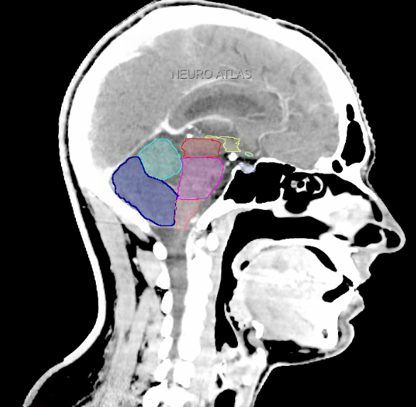
The European Particle Therapy Network (EPTN) has undertaken a major effort to harmonize OAR contouring practices in neuro-oncology. In 2018, EPTN published the first consensus-based guidelines for the delineation of 15 brain OARs (10.17195/candat.2017.08.1), expanded in 2021 to include 10 additional structures (10.17195/candat.2021.02.1). To aid adoption, EPTN also produced explanatory videos (10.17195/candat.2022.02.1), but the broad implementation of these guidelines remains limited due to the complexity and time-consuming contouring. In this context, deep learning (DL) offers a promising solution. Expert-trained DL models can encode complex anatomical knowledge and replicate expert-level contouring.

To this end, an nnUNet architecture has been trained and made public here. The model was trained using a 5-fold cross validation on 74 patients treated at Maastro Clinic, contoured according to the EPTN guidelines. The model receives as input a planning CT scan with intravenous iodine contrast, along with 3T MRI scan gadolinium-enhanced T1-weighted sequence. CT and MR image should be rigidly registered. The output of the model are the 25 OARs defined in the EPTN consensus guidelines.

EPTN International Neurological Contouring Atlas - 2021 Update

Daniëlle Eekers, Dario Di Perri, Erik Roelofs, Alida Postma, Esther Troost

Eekers et al. have published an international neurological atlas for contouring of organs at risk in consensus with the European Particle Therapy Network (EPTN) in 2018 and an update in 2021. The purpose of this consensus atlas is to decrease inter- and intra-observer variability in delineating OARs relevant for neuro-oncology.

Included are all OARs known to be relevant for radiation-induced toxicity in neuro-oncology: brain, brainstem (midbrain, pons, medulla oblongata), chiasm, cerebellum (anterior & posterior), cochlea, cornea, hippocampus (anterior & posterior), hypothalamus, lens, lacrimal gland, optic nerve, pituitary, skin, and vestibular & semicircular canals. To further facilitate research on cognition, vision and radiological changes after irradiation of the brain, potential clinically-relevant OARs are included: amygdala, caudate nucleus, cerebellum (anterior & posterior), corpus callosum, fornix, macula, optic tract, orbitofrontal cortex, periventricular space (PVS), pineal gland, and thalamus.

We propose this atlas is used in photon and particle therapy in order to derive consistent dosimetric data. When required this atlas will be updated according to new insights.

Three-dimensional delineation of the 25 consensus OARs for neuro-oncology are shown on CT (WW/WL 120/40, 3000/600), 3T MR images, (T1Gd, T2FLAIR 1mm) and 7T MR (MP2RAGE 0.7 mm). All are presented in transversal, sagittal and coronal view.

Please cite this Atlas as follows:

Eekers D, Di Perri D, Roelofs E, Postma A, Troost EG.
EPTN International Neurological Contouring Atlas - 2021 Update
CancerData, 2021. doi:10.17195/candat.2021.02.1


Optimization of Brain and Head & Neck Radiotherapy

Daniëlle BP Eekers

The aim of this thesis is to further optimise radiation therapy of Brain and Head & Neck cancer by reducing the dose to the healthy surrounding tissue, so called organs at risk (OARs), leading to a reduction in side effects.

Different treatment techniques for photon and particle (e.g. proton) therapy are compared for multiple tumour sites. Furthermore, tools for consistent contouring and dose prescription are presented in close collaboration with the European Particle Therapy Network (EPTN).


EPTN consensus-based guideline for the tolerance dose per fraction of organs at risk in the brain

Daniëlle BP Eekers, Maarten Lambrecht, Petra De Witt Nyström, Ans Swinnen, Frederik WR Wesseling, Erik Roelofs, Esther GC Troost


EPTN consensus-based guideline for the tolerance dose per fraction of organs at risk in the brain

In collaboration with the European Particle Therapy Network, a consensus table of tolerance doses for Organs at Risk in the brain was created. These values were all defined as a near maximum EQD2 values. Using the linear quadratic formula the doses are recalculated depending on the number of fractions.


Please site this table as follows:

Eekers D, Lambrecht M, Nyström PDW, Swinnen A, Wesseling FWR, Roelofs E, Troost EG
EPTN consensus-based guideline for the tolerance dose per fraction of organs at risk in the brain
CancerData, 2018; doi:10.17195/candat.2018.01.1

EPTN International Neurological Contouring Atlas

Daniëlle Eekers, Lieke In T Ven, Erik Roelofs, Alida Postma, Esther G Troost


Eekers et al. have published an international neurological atlas for contouring of organs at risk in consensus with the European Particle Therapy Network (EPTN). The purpose of this consensus atlas is to decrease inter- and intra-observer variability in delineating OARs relevant for neuro-oncology. We propose this atlas is used in photon and particle therapy in order to derive consistent dosimetric data. When required this atlas will be updated according to new insights.

Included are all OARs known to be relevant for radiation-induced toxicity in neuro-oncology: brain, brainstem, cochlea, vestibulum & semicircular canals, cornea, lens, retina, lacrimal gland, optic nerve, chiasm, pituitary, hippocampus and skin. A new OAR relevant for neuro-cognition, the posterior cerebellum is also included.

Three-dimensional delineation of the fifteen consensus OARs for neuro-oncology are shown on CT and 3 Tesla (3T) MR images (slice thickness 1 mm with intravenous contrast agent). All are presented in transversal, sagittal and coronal view.


Please cite this Atlas as follows:

Eekers D, In ’t Ven L, Roelofs E, Postma A, Troost EG. 
EPTN International Neurological Contouring Atlas 
CancerData, 2017. doi:10.17195/candat.2017.08.1